User guide
Instructions for using microcell arrays

1. Using a pair of tweezers lift the edge of the microcell array to peel the insert off the carrier slide. Always use gloves when working with the microcell arrays as contamination (e.g. dust, oils,...) from the skin can adversely affect the adhesive performance of the arrays.
2. Carefully place the microcell array into the imaging culture ware by laying the insert down from one edge to ensure that no air is trapped between the surface of the culture ware and the microcell array. If air bubbles become trapped, then remove the insert and try again. If the trapping of air bubbles continues, place one to two drops of ethanol onto the culture ware, then lay the insert down. Please note, if you use ethanol to assist in laying the insert down, leave the culture ware in the hood for at least 60 minutes to allow the ethanol to evaporate, otherwise the insert will not stick to the cell culture ware when the media is added.
NOTE: To check for trapped air bubbles under the array turn the culture ware upside down and look through the bottom surface.
3. The microcell array inserts are provided in vacuum sealed packaging; however, we recommend the following step for sterilization. Pipette 70 - 100% ethanol on top of the microcell array until the bottom surface of the culture ware including the insert is covered. Use a 1,000 μL pipette to cycle the ethanol in the culture ware in order to assist in removing any air bubbles that may be trapped in the wells. Finally use the pipette to draw out almost all the ethanol from the culture ware making sure that the array is always covered in liquid, and discard as waste.
NOTE: The microcell arrays are very hydrophobic and the addition of warm media to the arrays if they ethanol rinse is completely aspirated will result in air bubbles being trapped in the wells.
4. Slowly add warm media to one corner of the culture ware until almost full.
5. Draw out the media/ethanol mixture from the opposite corner of the culture ware, taking care that the microcell array is always covered with liquid.
6. Repeat steps 4 and 5 at least five times to make sure the ethanol is completely diluted out.
7. Pipette cells into the culture ware directly above the array. For a typical imaging experiment, seed between 1 - 2x10⁴ cells. Allow approximately 30 mins for the cells to settle before imaging.
User guide
Instructions for using microculture arrays - immunofluorescence assay

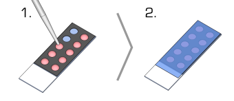
1. Place the microculture array into a 100mm culture dish and pipette the cell suspension directly into the isolated wells in the removable silicone array. NOTE: the array could also be removed from the glass slide and placed directly onto the surface of the culture dish, coverslip or other cell culture ware.
1 well - 250μL
2 well - 250μL
3 well - 200μL
10 well - 50μL
40 well - 8μL
2. Cover the culture dish with a lid to reduce evaporation and incubate the cells following standard procedures.
3. Remove the silicone array by gently lifting a corner with tweezers and peeling the array off the glass slide. NOTE: this step can be performed after fixing/staining.
4. Perform standard staining protocols on the slide (fixation, permeabilization, staining, washing).
5. Seal a coverslip (60 x 24mm) onto the glass slide with your choice of mounting medium.
NOTE: Typical well volume for pipetting cell suspension for cultivation.
Instructions for using microculture arrays - live cell imaging

1. Place the microculture array into a 100mm culture dish and pipette the cell suspension directly into the isolated wells in the removable silicone array. NOTE: the array could also be removed from the glass slide and placed directly onto the surface of the culture dish, coverslip or other cell culture ware.
1 well - 100μL
2 well - 100μL
3 well - 70μL
10 well - 20μL
40 well - 4μL
2. Seal the array with a coverslip (60 x 24mm) by gently pressing the coverslip down onto the array.
NOTE: Typical well volume for pipetting cell suspension for live cell imaging. Smaller medium volumes are required to prevent cross contamination when the array is sealed. The smaller liquid volume may not completely fill the well prior to sealing.
User guide
Protocol for poly-l-lysine coating a microcell array
Materials
Either poly-lysine or poly-D-lysine with MW between 30,000 - 150,000 can be used for the coating.
Prepare 1 mg/mL stock solution by dissolving 100 mg of poly-D-lysine hydrobromide in 100 mL of PBS. The stock solution can be stored at -20°C.
The working solution is obtained by diluting the stock solution with PBS to around 0.1 mg/mL.
Procedure
1. Insert the microcell array into the chamber to be used for imaging.
2. Using a 1,000 μL pipette slowly add 500 μL of 100% ethanol on top of the microcell array, then cycle the ethanol over the microcell array to assist in removing any trapped air bubbles.
3. Slowly add media to one corner of the imaging chamber until almost full.
4. Draw out the media/ethanol mixture from the opposite corner of the imaging chamber, taking care that the microcell array is always covered with a small amount of liquid. Repeat steps 2 and 3 at least five times to make sure that the ethanol is completely diluted out.
5. Add the working solution of poly-lysine to the microcell array.
6. Incubate for 1 hour at room temperature in a hood.
7. Carefully aspirate the poly-lysine solution from the imaging chamber, taking care that the microcell array is always covered with a small amount of liquid. Note that the poly-lysine working solution can be re-used a few times.
8. Rinse 3 times with PBS, taking care that the microcell array is always covered with a small amount of liquid.
9. The poly-lysine coated microcell array should be used immediately.
Protocol for collagen coating a microcell array
Materials
Rat tail collagen (type 1) can be used for the coating. It is recommended that you purchase collagen solutions.
Dilute the purchased solution to 50 μg/mL with 0.02M acetic acid to produce the working solution.
Procedure
1. Insert the microcell array into the chamber to be used for imaging.
2. Using a 1,000 μL pipette slowly add 500 μL of 100% ethanol on top of the microcell array, then cycle the ethanol over the microcell array to assist in removing any trapped air bubbles.
3. Slowly add media to one corner of the imaging chamber until almost full.
4. Draw out the media/ethanol mixture from the opposite corner of the imaging chamber, taking care that the microcell array is always covered with a small amount of liquid. Repeat steps 2 and 3 at least five times to make sure that the ethanol is completely diluted out.
5. Add the working solution of collagen (50 μg/mL) to the microcell array at 5 μg/cm2.
6. Incubate for 1 hour at room temperature in a hood.
7. Carefully aspirate the collagen solution from the imaging chamber, taking care that the microcell array is always covered with a small amount of liquid.
8. Rinse 3 times with PBS, taking care that the microcell array is always covered with a small amount of liquid.
9. The collagen coated microcell array should be used immediately.
Protocol for fibronectin coating a microcell array
Materials
Human fibronectin is diluted to 10 - 50 μg/mL with Ca++ and Mg++ free PBS to produce the working solution. Note that the concentration should be proportional to the coating concentration. For example, make 10 μg/mL solution for 1 μg/cm2 coating and 50 μg/mL solution for 5 μg/cm2 coating.
Make appropriate aliqouts and store at -20°C and avoid multiple freeze/thaw cycles.
Procedure
1. Insert the microcell array into the chamber to be used for imaging.
2. Using a 1,000 μL pipette slowly add 500 μL of 100% ethanol on top of the microcell array, then cycle the ethanol over the microcell array to assist in removing any trapped air bubbles.
3. Slowly add media to one corner of the imaging chamber until almost full.
4. Draw out the media/ethanol mixture from the opposite corner of the imaging chamber, taking care that the microcell array is always covered with a small amount of liquid. Repeat steps 2 and 3 at least five times to make sure that the ethanol is completely diluted out.
5. Add the appropriate amount of fibronectin solution to the microcell array.
6. Incubate for 1 hour at room temperature in a hood.
7. Carefully aspirate the fibronectin solution from the imaging chamber, taking care that the microcell array is always covered with a small amount of liquid.
8. Rinse 3 times with PBS, taking care that the microcell array is always covered with a small amount of liquid.
9. The fibronectin coated microcell array should be used immediately.
Protocol for laminin coating a microcell array
Materials
Laminin is diluted to 5 μg/mL in PBS. The concentration should be proportional to the coating concentration. For example, make 10 μg/mL solution for 1 μg/cm2 coating and 50 μg/mL solution for 5 μg/cm2 coating.
Make appropriate aliqouts and store at -20°C and avoid multiple freeze/thaw cycles.
Procedure
1. Insert the microcell array into the chamber to be used for imaging.
2. Using a 1,000 μL pipette slowly add 500 μL of 100% ethanol on top of the microcell array, then cycle the ethanol over the microcell array to assist in removing any trapped air bubbles.
3. Slowly add media to one corner of the imaging chamber until almost full.
4. Draw out the media/ethanol mixture from the opposite corner of the imaging chamber, taking care that the microcell array is always covered with a small amount of liquid. Repeat steps 2 and 3 at least five times to make sure that the ethanol is completely diluted out.
5. Add an appropriate amount of laminin solution to the microcell array.
6. Incubate for 1 hour at room temperature in a hood.
7. Carefully aspirate the laminin solution from the imaging chamber, taking care that the microcell array is always covered with a small amount of liquid.
8. Rinse 3 times with PBS, taking care that the microcell array is always covered with a small amount of liquid.
9. The Laminin coated microcell array should be used immediately.
Microcell array not sticking to the imaging chamber surface
1. Inspect the quality of the insert's seal against the cell cutlure ware by looking through the bottom of the cell culture ware. The presence of air bubbles will reduce the sticking performance of the insert. Remove the insert and try again.
2. If no air bubbles are present and the insert will not stick to the cell culture ware surface, then remove the insert from the cell culture ware.
3. Carefully place the bottom surface of the microcell array onto some 3M Scotch Magic tape. The insert can aggresively collect dust/oils that reduces its ability to self-adhere to a surface. The scotch tape removes the dust/oils enabling the insert to adhere to the surface of the imaging chamber.
NOTE: general sticky tape will not work as it can leave glue residue on the insert.
4. Using a pair of tweezers carefully peel the insert off the tape and place it onto a clean section of tape.
5. Repeat steps 3 and 4 at least 3 times.
6. Return the microcell array to the cell culture ware. Make sure that the insert is only ever applied to a dry surface.
NOTE: if the microcell array is still not sticking to the surface of the imaging chamber then after step 6 place the imaging chamber on a hot plate at 60°C for 10min.


Permanently bonding a microcell array to a glass surface
1. Clean the insert and glass surface of the imaging chamber in ethanol and dry with compressed air or nitrogen.
NOTE: the inserts will only bond with a glass surface.
2. Activate the surfaces of the insert and imaging chamber using an Oxygen plasma or corona discharge for approximately 15 seconds.
3. Carefully place the insert onto the glass surface and press down on all areas of the insert making sure it is in contact with the glass.
NOTE: if you have made a mistake or a bubble has become trapped between the insert and glass surface you only have one to two minutes to remove the insert and try again before it bonds with the glass.
4. Apply pressure to the insert using your finger for 30 seconds.
5. Wait at least 1 hour before starting the ethanol rinse (step 3 in the protocol for using microcell arrays).

Matlab tool box for processing time lapse movies of cells
John Markham from the National ICT Australia (NICTA) has developed a MATLAB based tool box called MATS (Microgrid Array Tools) to process time lapse microscopy experiments using microgrid arrays. Unlike other processing software MATS has some specific features for use with microgrid arrays like;
1. Automatic recognition of microgrid boudaries. There are three algorithms which can be used with transmission images to find the microgrid boundaries.
2. Support for spectral unmixing based on the Zeiss Colibri illumination with a quad-band filter block.
3. Support for automatic position setting. Setting positions that cover all of the 1,000's of wells in a microgrid array is a painful task, so a script is included that automates it, including doing interpolation of the focus to account for a non-level holder.
4. Support for measurement of FUCCI cell cycle reporter proteins.
5. Support for multi-treading and GPU acceleration, which allows you to use as many cores on your local machine as the MATLAB parallel tool box will support.
6. Generation of position specific background images. MATS uses all the images in a stack to try and find the background. THis inly works if the cells are motile and take up only a small proportion of the image.
7. Support for image registration to account for misalignment at successive time points.

